If you’re preparing for the United States Medical Licensing Examination® (USMLE®) Step 1 exam, you might want to know which questions are most often missed by test takers. Check out this example from Kaplan Medical, and view an expert video explanation of the answer. Also check out all posts in this series.
This month’s stumper
A 41-year-old woman comes to the physician because of a recent history of joint pain in her hands and knees, in addition to some mild weight loss and fatigue. She has a two-week history of swelling behind her left knee. She states that the swelling has been progressively growing, and she has pain and difficulty in flexing the knee. Her serum is positive for rheumatoid factor. Physical examination shows an erythematous, fluid-filled mass in the left lateral popliteal fossa overlying the tendon of the biceps femoris. During aspiration of the cyst, a structure immediately adjacent to the tendon is injured.
Which of the following losses is most likely to occur in this patient?
A. Loss of dorsiflexion at the ankle
B. Loss of flexion of the toes
C. Loss of plantar flexion at the ankle
D. Loss of sensation on the sole of the foot
E. Vascular insufficiency to the leg and foot
The correct answer is A.
Kaplan Medical explains why
This patient presents with a Baker's (popliteal) cyst, a cystic enlargement of the popliteal bursa. It is filled with synovial fluid. The area can become inflamed and is therefore painful. Depending on cyst size, there may be limitation of movement at the knee, especially in flexion. Such cysts are often associated with rheumatoid arthritis. Most of these cysts are small and resolve spontaneously; however, if the cyst is significant, as in this question, needle aspiration is usually performed.
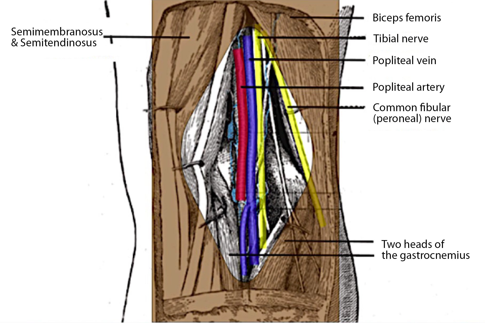
The popliteal fossa is a diamond-shaped region behind the knee that is bounded by:
- The tendon of the biceps femoris, superolaterally
- The tendons of the semimembranosus and semitendinosus, superomedially
- The two heads of the gastrocnemius, inferolaterally and inferomedially
In the lateral part of the fossa, the common fibular (peroneal) nerve lies immediately adjacent to the tendon of the biceps femoris and might be injured in this procedure. The common fibular nerve divides into the superficial fibular and deep fibular nerves. The deep fibular nerve innervates the muscles of the anterior compartment of the leg (tibialis anterior, extensor hallucis longus and extensor digitorum). These muscles are all dorsiflexors of the ankle.
Why the other answers are wrong
Read these explanations to understand the important rationale for each answer.
Choice B: Flexion of the toes is caused by the flexor digitorum and flexor hallucis muscles. These muscles are supplied by the tibial nerve, which passes through the center of the popliteal fossa and is not likely to be injured during this aspiration.
Choice C: The plantar flexors of the ankle are the muscles of the posterior compartment of the leg. These muscles are innervated by the tibial nerve, which passes through the center of the popliteal fossa and is not likely to be injured during this aspiration.
Choice D: Sensation on the sole of the foot is supplied by the medial and lateral plantar nerves, which are both branches of the tibial nerve, which, as indicated above, is not likely to be injured during this aspiration.
Choice E: The vascular supply distal to the knee is from the popliteal artery. The popliteal artery passes through the center of the popliteal fossa and is very deeply placed in the fossa and not likely to be injured during this aspiration.
Tips to remember
- The structures that border the popliteal fossa superiorly are the biceps femoris laterally and the semimembranosus and semitendinosus medially.
- Inferiorly, the gastrocnemius muscle borders the fossa medially and laterally.
- The tibial nerve, popliteal vein and popliteal artery course inferiorly through the middle of the fossa, and the common fibular nerve courses inferolaterally, adjacent to the biceps femoris tendon.
For more prep questions on USMLE Steps 1 and 2, view other posts in this series.


