If you’re preparing for the United States Medical Licensing Examination (USMLE) Step 1 exam, you might want to know which questions are most often missed by test-prep takers. Check out this example from Kaplan Medical, and read an expert explanation of the answer. Also check out all posts in this series.
This month’s stumper
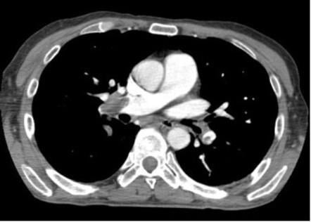
A 66-year-old man is brought to the emergency department after recent discharge following a Whipple’s procedure for pancreatic cancer performed seven days prior. He has a six-hour history of worsening shortness of breath and sudden onset chest pain. He is given oxygen supplementation, which moderately improves his saturation. A contrast-enhanced CT scan of the chest is shown. Which of the following is the most likely origin of the abnormality seen on CT?
A. Basilic vein
B. Brachial vein
C. Cephalic vein
D. Femoral vein
E. Great saphenous vein
F. Lesser saphenous vein
The correct answer is D.
Kaplan Medical explains why
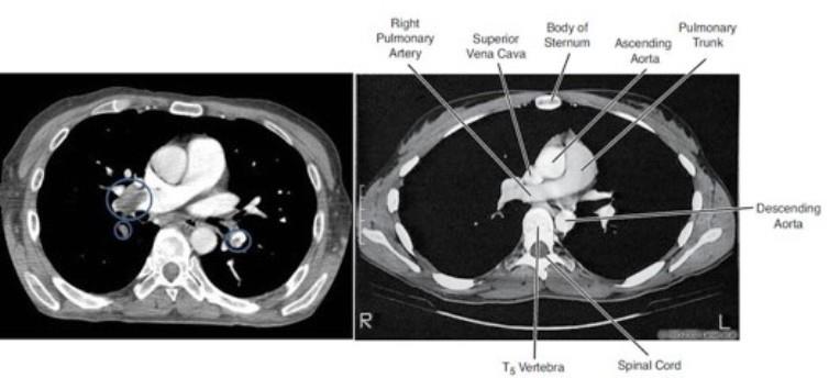
The patient most likely has a pulmonary embolism (PE). The large filling defect in the right pulmonary artery and in the superior branch of the left pulmonary artery (see circles in the left image below) in the CT scan support the diagnosis. For comparison, a normal image through the same region is shown on the right.
More than 90 percent of pulmonary emboli originate from the deep veins of the lower limbs. The only deep vein of the lower limb listed in the answer choices is the femoral vein. Venous thromboses can also form more distally in the popliteal vein. The risk of embolism increases as the clot extends proximally.
Why you shouldn’t choose the other answers
Read these explanations to understand the important rationale for each answer.
Choices A and C: The basilic and cephalic veins are two superficial veins of the upper limbs and are the ones most frequently accessed with IV catheters. Virtually the only time you see major clots in the upper extremities is when they contain some sort of catheter.
Choice B: The brachial vein is the major deep vein of the upper extremities. Venous thromboses can form in the deep veins of the upper extremities but venous thromboembolism much more commonly arises from DVTs in the lower extremities.
Choices E and F: The great saphenous and lesser saphenous veins are the superficial veins of the lower limbs and do not commonly result in pulmonary embolism. The great saphenous vein is commonly harvested for bypass procedures.
Key tips to remember
- More than 90 percent of pulmonary emboli begin in the deep veins of the lower limbs.
- Deep vein thromboses of the lower extremities generally form in the popliteal veins and extend proximally.
- The more proximal the clot, the more likely it is to embolize to the lungs.
For more prep questions on USMLE Steps 1, 2 and 3, view other posts in this series.
The AMA and Kaplan have teamed up to support you in reaching your goal of passing the USMLE® or COMLEX-USA®. If you're looking for additional resources, Kaplan provides free access to tools for pre-clinical studies, including Kaplan’s Lecture Notes series, Integrated Vignettes, Shelf Prep and more.


